
Hero Image
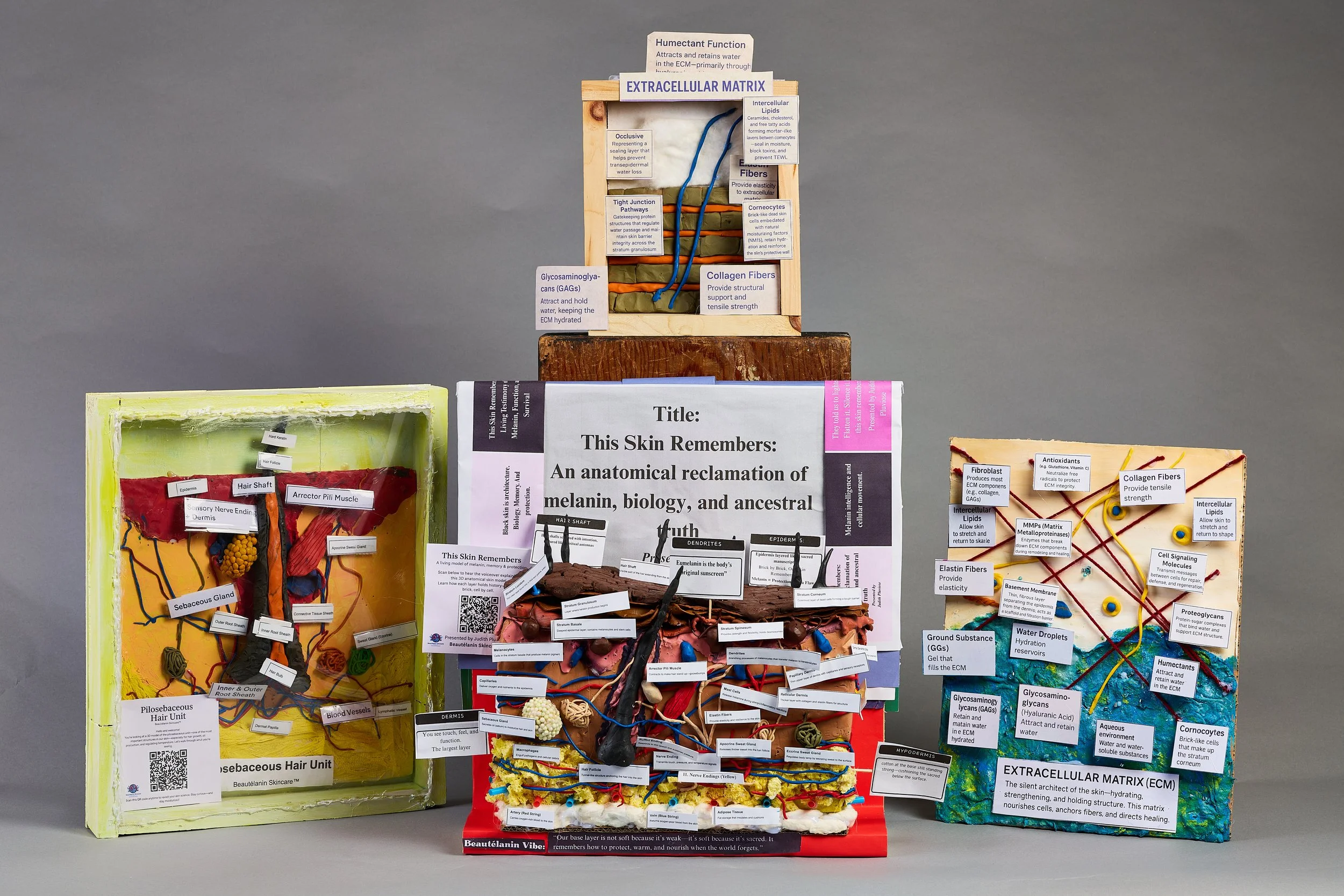
Full Installation View
All three models together
sc_0127.jpg
Educational Art & Anatomical Models · Beautélanin™
Handmade, three-dimensional anatomical models built to make skin science visible, accessible, and culturally relevant — for students, practitioners, educators, and communities who were never centered in the conversation.
Every model began with a question education forgot to answer.
These are not diagrams. They are objects you can study, touch, and return to — built to carry scientific knowledge in a form that textbooks rarely achieve.
Each model maps a different layer of the skin's architecture: from the surface epidermis and its dendrites, through the layered dermis and its vessels and glands, to the extracellular matrix and the fat-cushioned hypodermis beneath. All labeled. All explained. All made by hand.
Black skin is architecture. Biology, memory, and protection — built into every layer.
Featured Collection · 01
An Anatomical Reclamation of Melanin, Biology, and Ancestral Truth
Main Installation Photo
Front View with Labels
sc_0017.jpg
Project Statement
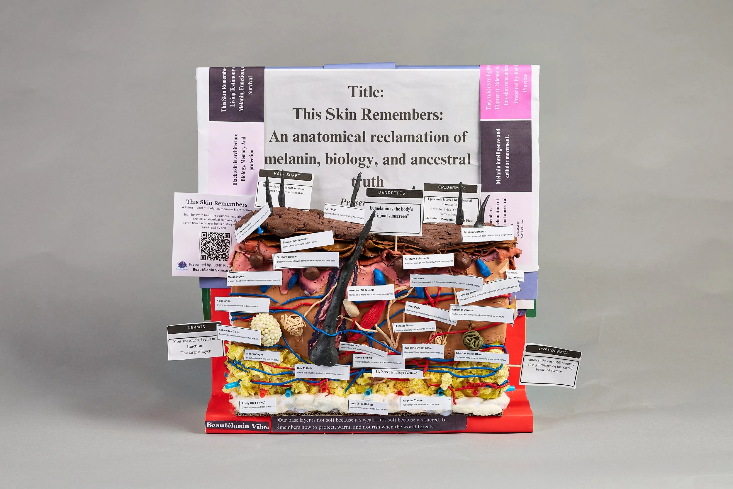
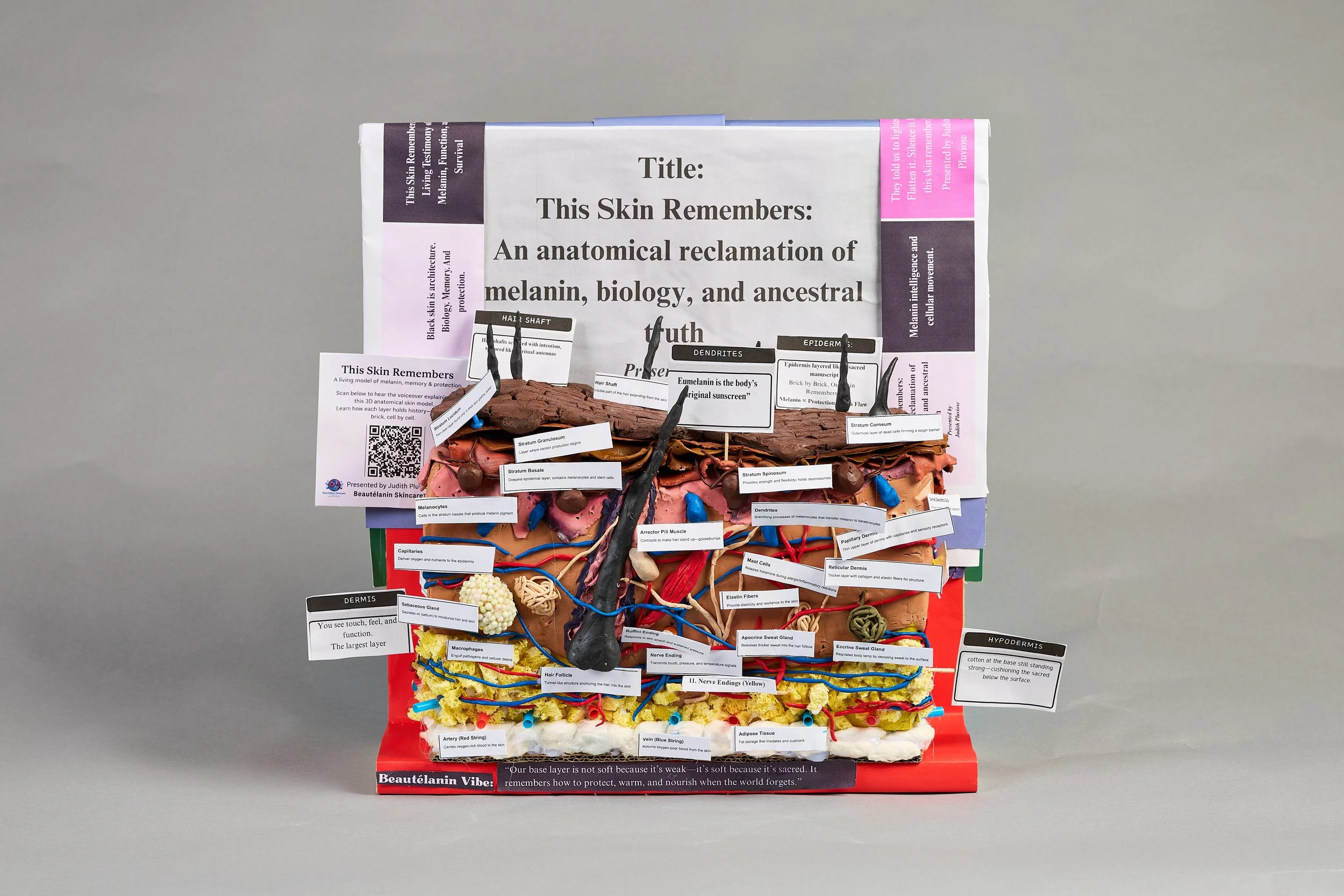
A 3D model of melanin, cellular architecture, and protection — constructed across more than 30 anatomical structures, cell by cell, layer by layer.
The model is presented in cross-section to make the invisible visible: to show what skin is, not simply what it looks like.
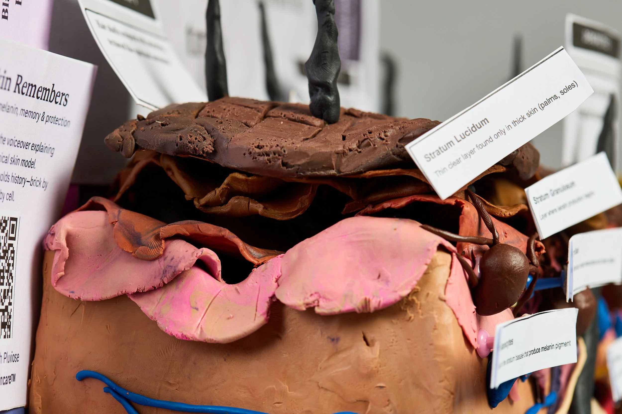
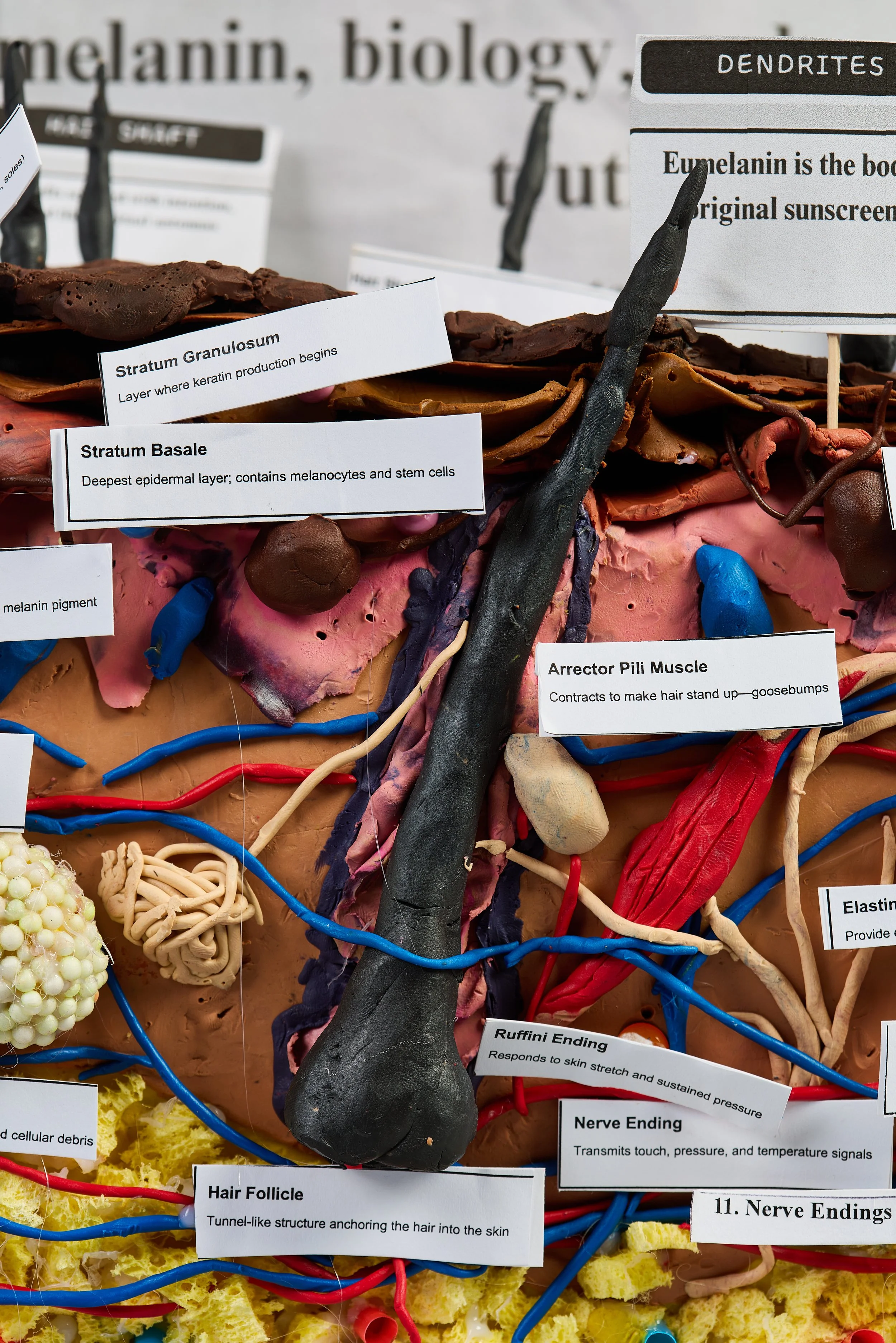
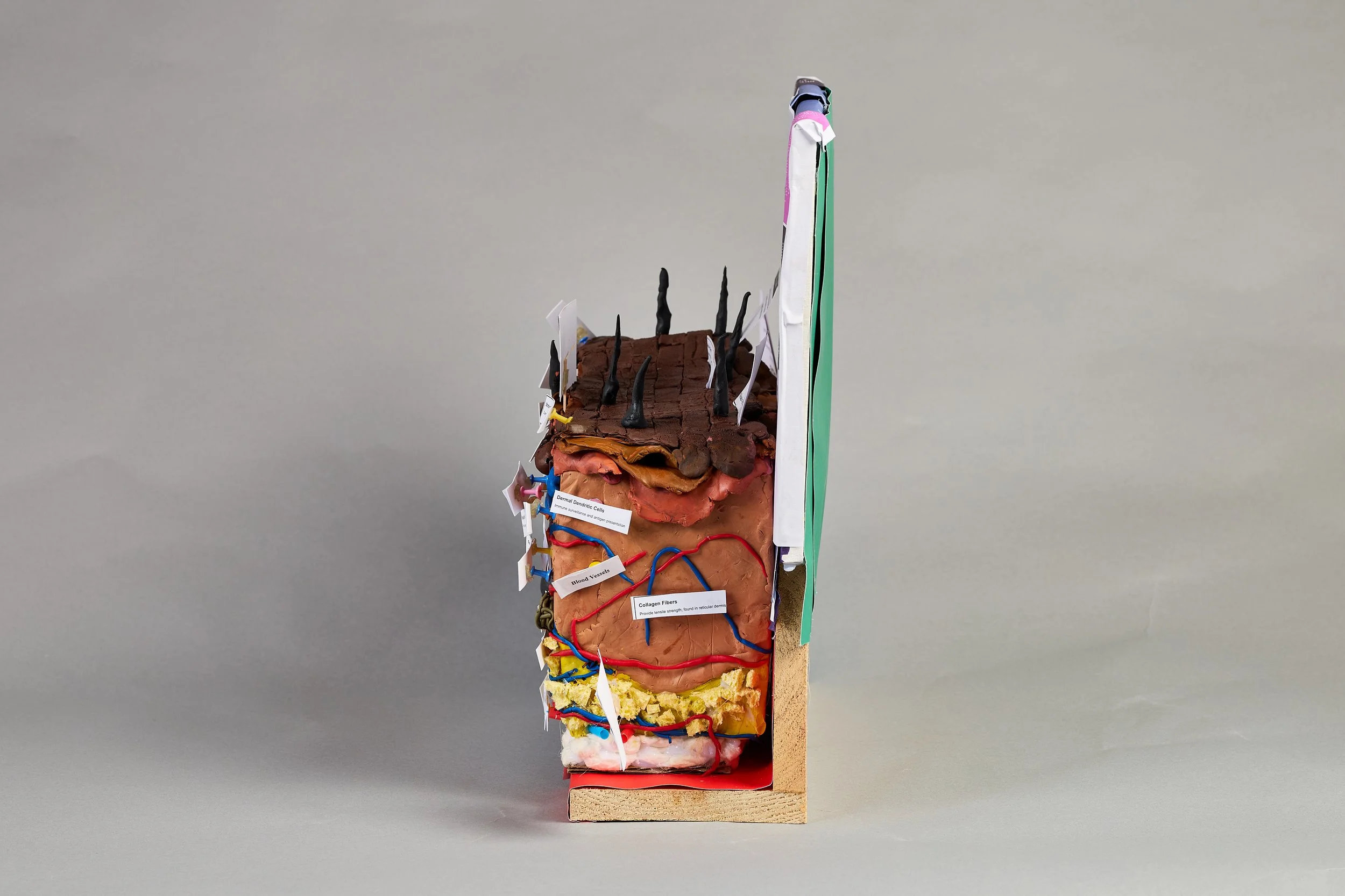
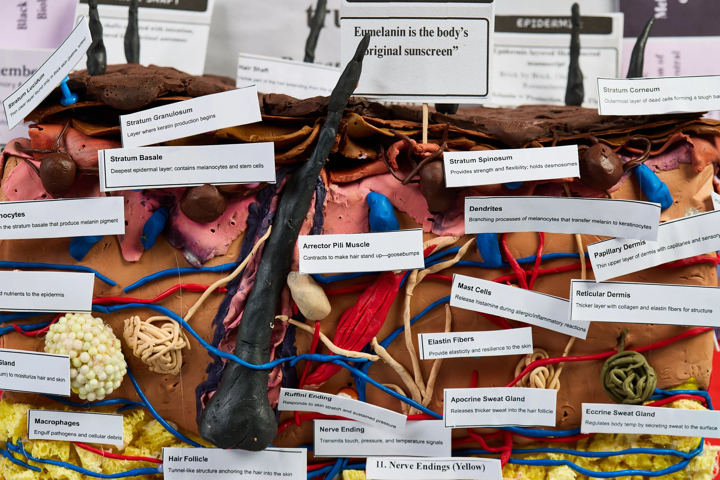
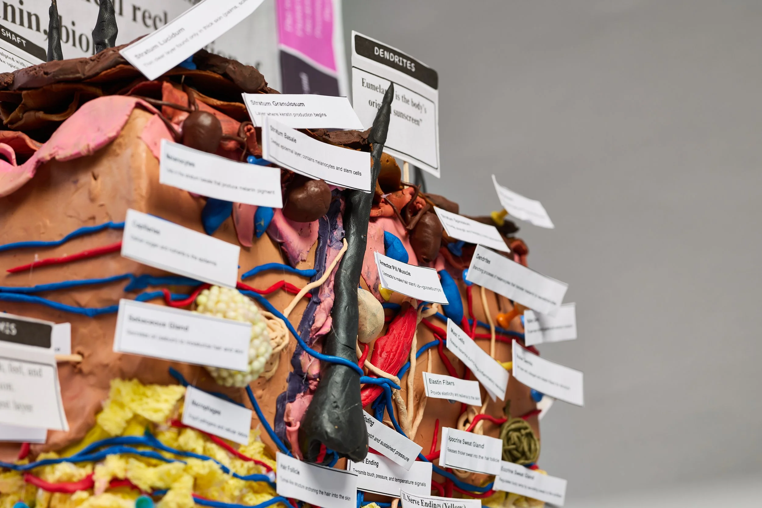
Beginning at the stratum corneum — where corneocytes stack like brick and mortar — the model moves through each epidermal layer: granulosum, spinosum, basale. Melanocytes sit at the base, extending their dendrites upward. Eumelanin is the body's original sunscreen.
Below the epidermis, the dermis holds the model's richest detail: arrector pili muscle, sebaceous and apocrine glands, elastic and collagen fibers, capillaries running red, veins returning blue, mast cells, macrophages, nerve endings in yellow.
At the base: the hypodermis. Adipose tissue — not fat as flaw, but fat as cushion, insulation, protection. Sacred beneath the surface.
"Our base layer is not soft because it's weak — it's soft because it's sacred. It remembers how to protect, warm, and nourish when the world forgets."
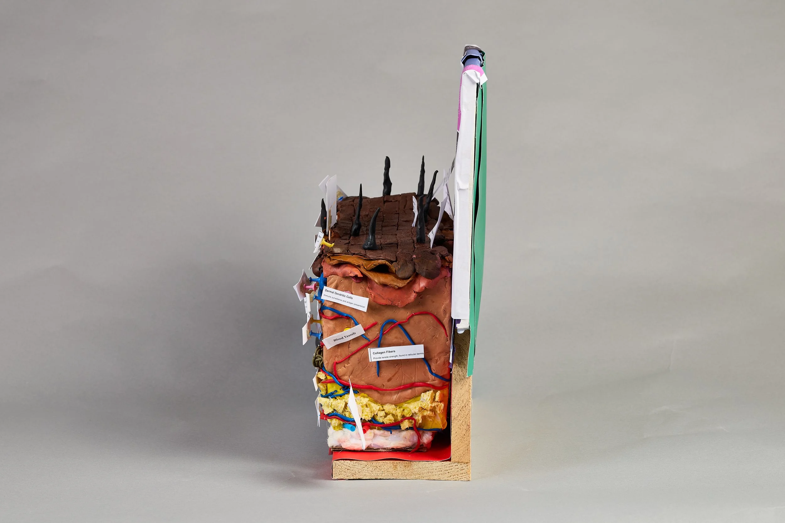
Side View — Right
Dermis & Layer Depth
sc_0019.jpg
Side View — Left
Layer Architecture
sc_0023.jpg
Detail Close-Up
Dendrites · Melanocytes · Arrector Pili
sc_0024.jpg
Presented by Judith S. Pluviose, Beautélanin™ Skincare. This model includes a QR code linking to an audio voiceover walkthrough, allowing viewers to hear the science as they study each layer. Every structure is labeled with its anatomical name and function. The goal: to make anatomy feel like testimony — not like a textbook.
Collection · 02
The Silent Architect of the Skin
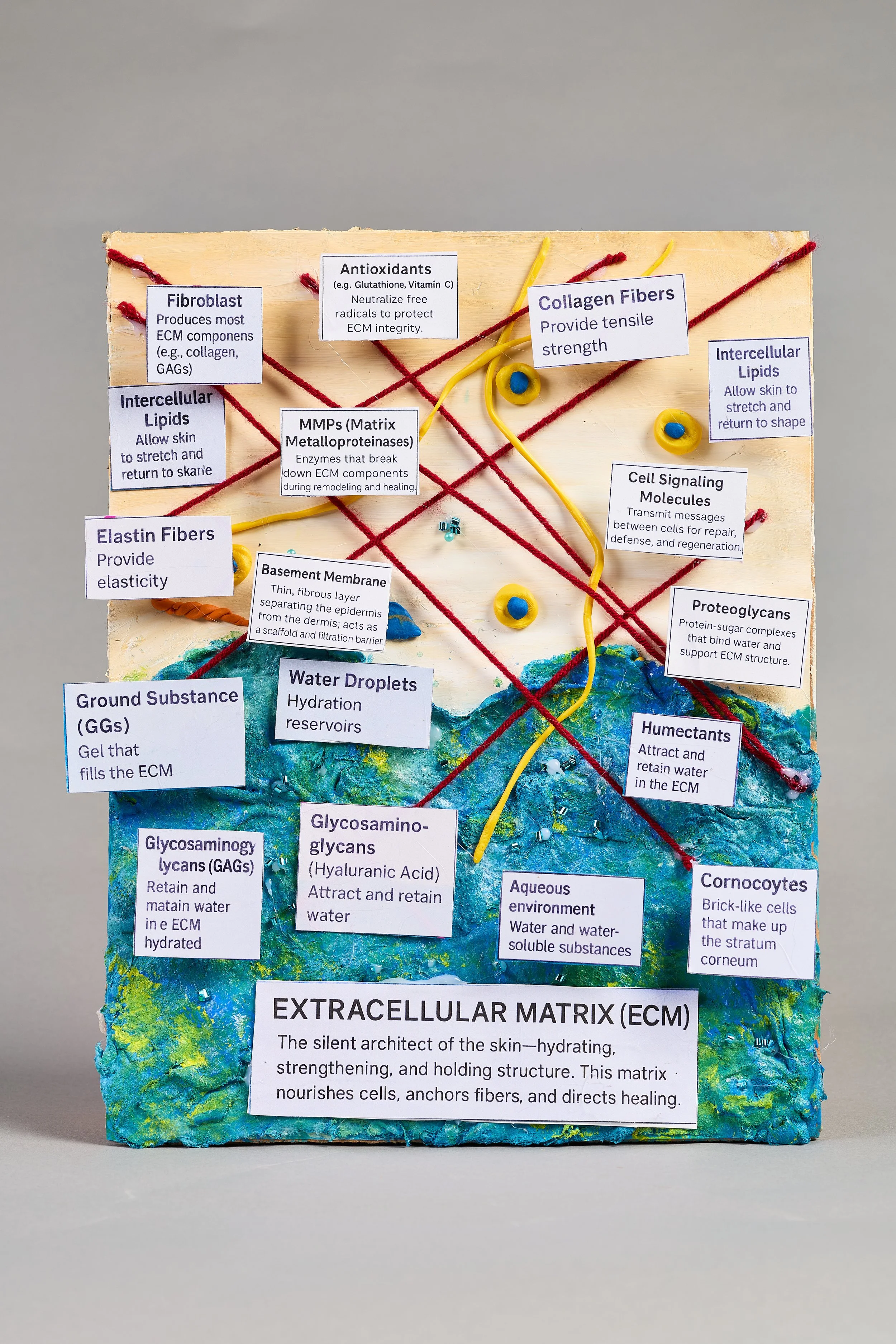
ECM Panel — Main View
Full Labeled Components
sc_0082.jpg
Educational Purpose
A tactile representation of the extracellular matrix — the gel-like scaffolding that surrounds every cell in the skin's dermis and epidermis.
Most skincare education skips the ECM entirely. This model makes it impossible to overlook.
The model is divided into two zones. The upper panel represents the dermal ECM: collagen fibers (red yarn), elastin fibers (yellow), water droplets (blue clay), fibroblasts, and the basement membrane — the thin layer separating dermis from epidermis.
The lower blue-green zone represents the aqueous ECM environment: glycosaminoglycans (GAGs), hyaluronic acid, corneocytes, and the aqueous medium that nourishes every cellular component.
"The ECM is the silent architect of the skin — hydrating, strengthening, and holding structure. This matrix nourishes cells, anchors fibers, and directs healing."
Barrier ECM Model
Wooden Shadowbox Frame
sc_0071.jpg
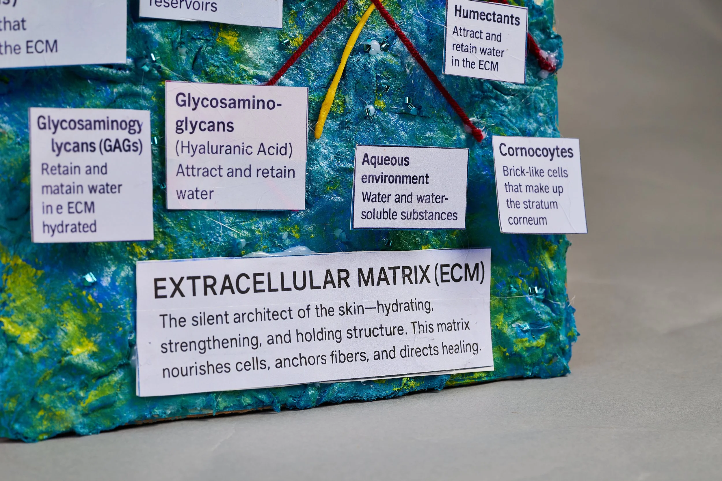
ECM Panel Detail
Macro Shot / Alternate Angle
Additional ECM detail photo
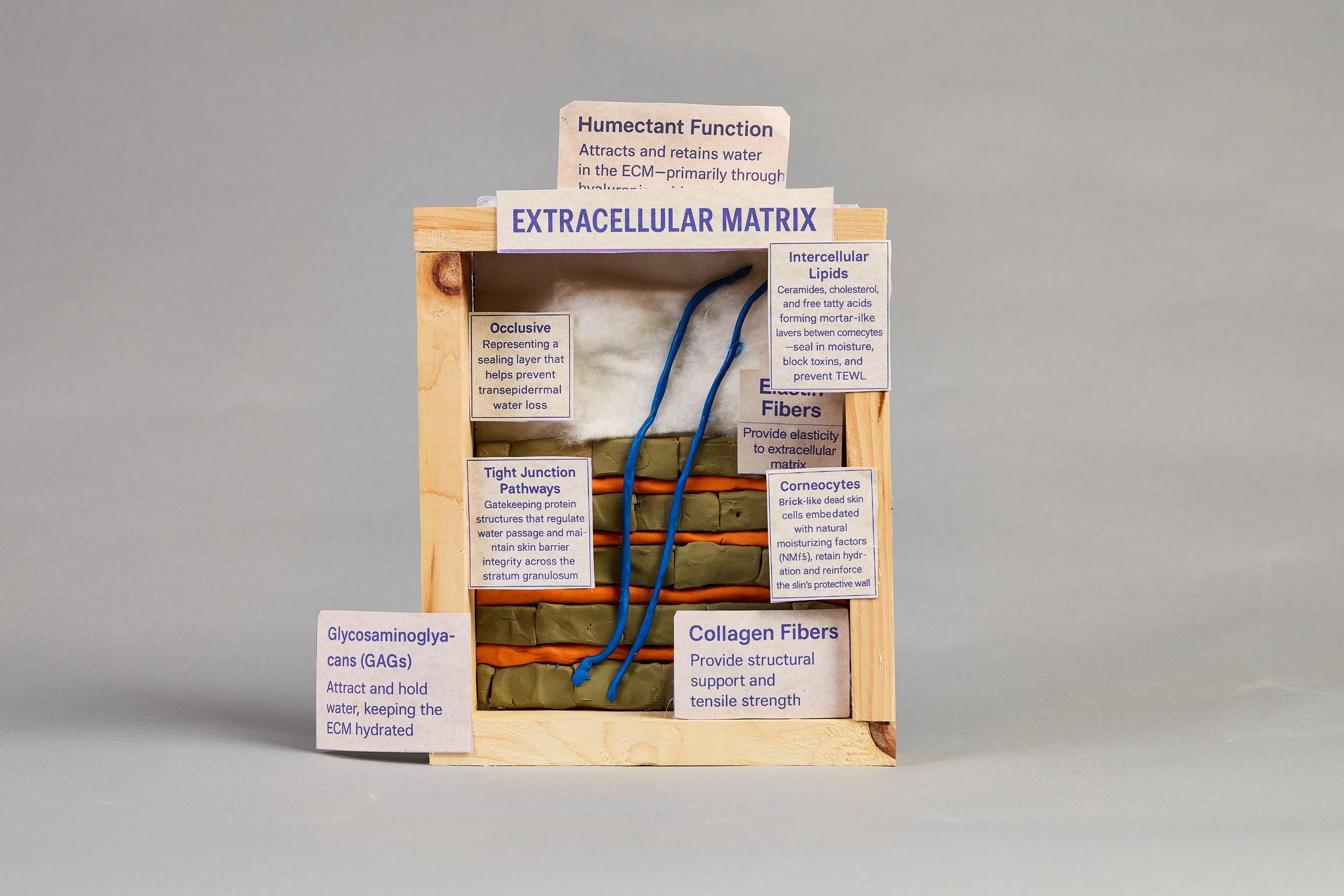
The barrier ECM shadowbox maps barrier-specific components: corneocytes, intercellular lipids, tight junction pathways, occlusive layer, elastin fibers, and GAGs — all annotated. Together with the larger ECM panel, these two models form a complete picture of how the skin holds, communicates, and repairs itself at the molecular level.
Collection · 03
Making Complex Skin Anatomy Accessible
Pilosebaceous Hair Unit
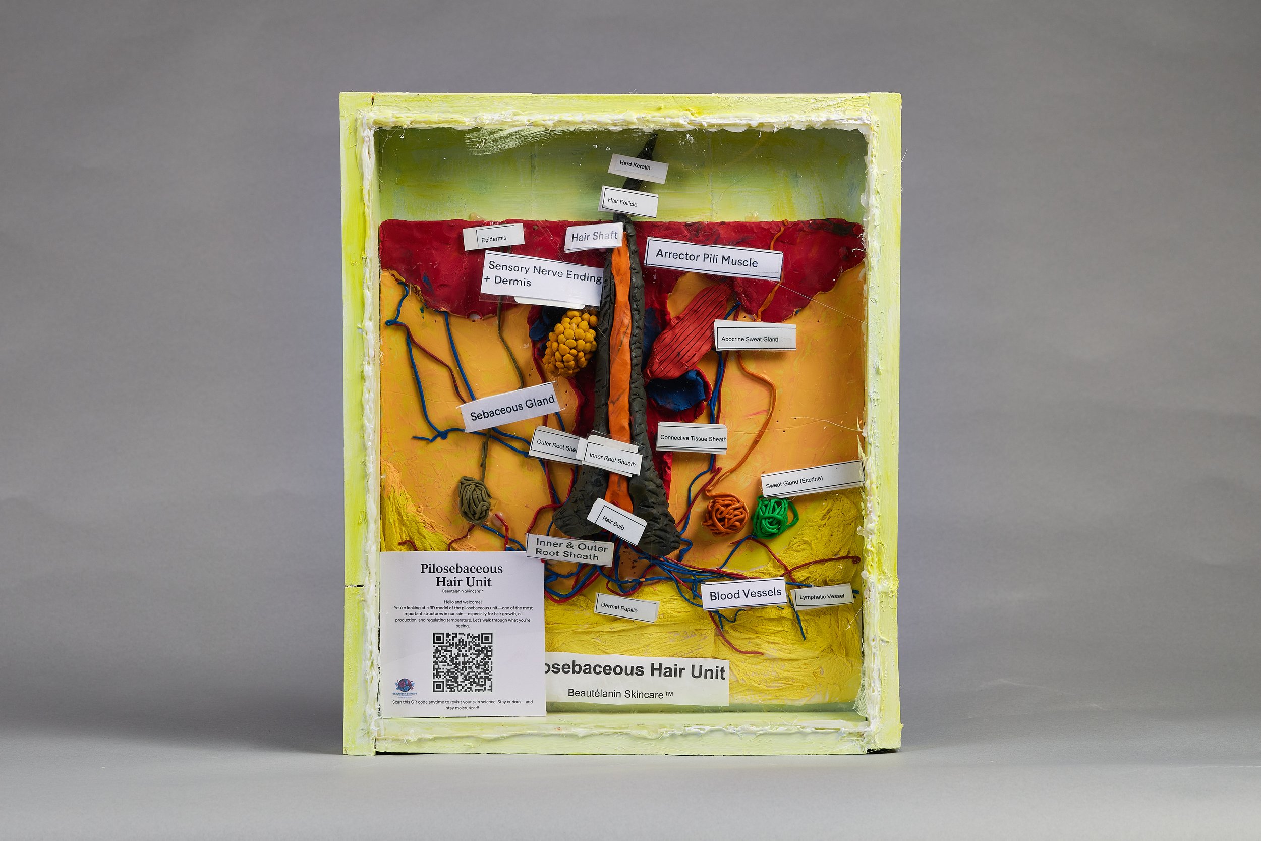
Full Shadowbox View
sc_0104.jpg
Medium
A three-dimensional interpretation of the pilosebaceous unit — one of the most important structures in the skin, especially for hair growth, oil production, and temperature regulation.
Built inside a framed shadowbox with a clear viewing face so the interior architecture is fully visible from the front.
The model maps the complete pilosebaceous system: the hair shaft descending into the follicle and dermal papilla; the arrector pili muscle (the structure that causes goosebumps); the sebaceous gland and its connection to the follicle; inner and outer root sheaths; apocrine and eccrine sweat glands; sensory nerve endings; and blood and lymphatic vessels that supply the entire unit.
Surrounding skin layers are clearly distinguished: epidermis (red clay) across the top; dermis (yellow/orange) through the middle zone; hypodermis at the base.
"You're looking at one of the most important structures in our skin — especially for hair growth, oil production, and regulating temperature. Let's walk through what you're seeing."
Detail
Hair Shaft & Follicle
Macro shot of hair structure
Detail
Sebaceous & Sweat Glands
Macro shot of gland structures
Detail
Blood Vessels & Nerve Endings
Macro shot of vascular structures
This model includes a QR code linking to a guided audio walkthrough. Every structure is labeled with its anatomical name and a one-sentence function summary, making it appropriate for clients, students, and practitioners at every level of training. The yellow shadowbox format allows the model to stand upright as a self-teaching display in a classroom, treatment room, or exhibition setting.
A personal statement from Judith S. Pluviose, founder of Beautélanin™.
Licensed Esthetician
Attorney
Certified Skincare Formulator
Educator
Founder, Beautélanin™
I began building anatomical models because I wanted students to see skin differently — not as defects to correct, but as living systems with history, function, and intelligence.
Most skincare education introduces anatomy through flat diagrams, briefly, before moving on to products and procedures. The skin becomes a surface to be managed rather than a system to be understood.
I wanted to make the skin impossible to dismiss. When you can hold a cross-section in your hands — when you can trace the path of a nerve ending or follow a blood vessel from artery to vein — the skin stops being abstract. It becomes real. It becomes remarkable.
Every model is handmade and designed to make anatomy visible, accessible, and culturally relevant.
These models were built with melanin-rich skin in mind from the beginning — centering eumelanin, post-inflammatory pathways, barrier considerations, and the biological realities that textbooks so often leave out. Because what you see shapes what you believe. And what you believe shapes how you treat people.
Where This Work Belongs
These are not classroom props. They are exhibition-quality objects that belong in spaces where science, culture, and community intersect.
Black History Museums
Centering Black anatomy, biology, and science as cultural heritage
Community Cultural Centers
Making skin science accessible and communally owned
Public Libraries
Free, open educational access to anatomical knowledge
University Diversity Programs
Supporting inclusive STEM and healthcare curricula
Medical Humanities Exhibits
Bridging science, art, and lived human experience
Public Health Exhibitions
Building community health literacy from the ground up
Skin-of-Color Conferences
Supporting practitioners and researchers in dermatology and esthetics
Esthetics Schools & Programs
Giving students what textbooks rarely provide
Continuing Education Conferences
Advanced visual learning for licensed practitioners
BEAUTÉLANIN™
The skin is not a flaw to be corrected.
It is a system to be understood.
Rooted in Haitian Heritage. Powered by NoirScience™.
Hero Image
Full Installation View
All three models together
sc_0127.jpg
Educational Art & Anatomical Models · Beautélanin™
Handmade, three-dimensional anatomical models built to make skin science visible, accessible, and culturally relevant — for students, practitioners, educators, and communities who were never centered in the conversation.
Every model began with a question education forgot to answer.
These are not diagrams. They are objects you can study, touch, and return to — built to carry scientific knowledge in a form that textbooks rarely achieve.
Each model maps a different layer of the skin's architecture: from the surface epidermis and its dendrites, through the layered dermis and its vessels and glands, to the extracellular matrix and the fat-cushioned hypodermis beneath. All labeled. All explained. All made by hand.
Black skin is architecture. Biology, memory, and protection — built into every layer.
Featured Collection · 01
An Anatomical Reclamation of Melanin, Biology, and Ancestral Truth
Main Installation Photo
Front View with Labels
sc_0017.jpg
Project Statement
A 3D model of melanin, cellular architecture, and protection — constructed across more than 30 anatomical structures, cell by cell, layer by layer.
The model is presented in cross-section to make the invisible visible: to show what skin is, not simply what it looks like.
Beginning at the stratum corneum — where corneocytes stack like brick and mortar — the model moves through each epidermal layer: granulosum, spinosum, basale. Melanocytes sit at the base, extending their dendrites upward. Eumelanin is the body's original sunscreen.
Below the epidermis, the dermis holds the model's richest detail: arrector pili muscle, sebaceous and apocrine glands, elastic and collagen fibers, capillaries running red, veins returning blue, mast cells, macrophages, nerve endings in yellow.
At the base: the hypodermis. Adipose tissue — not fat as flaw, but fat as cushion, insulation, protection. Sacred beneath the surface.
"Our base layer is not soft because it's weak — it's soft because it's sacred. It remembers how to protect, warm, and nourish when the world forgets."
Side View — Right
Dermis & Layer Depth
sc_0019.jpg
Side View — Left
Layer Architecture
sc_0023.jpg
Detail Close-Up
Dendrites · Melanocytes · Arrector Pili
sc_0024.jpg
Presented by Judith S. Pluviose, Beautélanin™ Skincare. This model includes a QR code linking to an audio voiceover walkthrough, allowing viewers to hear the science as they study each layer. Every structure is labeled with its anatomical name and function. The goal: to make anatomy feel like testimony — not like a textbook.
Collection · 02
The Silent Architect of the Skin
ECM Panel — Main View
Full Labeled Components
sc_0082.jpg
Educational Purpose
A tactile representation of the extracellular matrix — the gel-like scaffolding that surrounds every cell in the skin's dermis and epidermis.
Most skincare education skips the ECM entirely. This model makes it impossible to overlook.
The model is divided into two zones. The upper panel represents the dermal ECM: collagen fibers (red yarn), elastin fibers (yellow), water droplets (blue clay), fibroblasts, and the basement membrane — the thin layer separating dermis from epidermis.
The lower blue-green zone represents the aqueous ECM environment: glycosaminoglycans (GAGs), hyaluronic acid, corneocytes, and the aqueous medium that nourishes every cellular component.
"The ECM is the silent architect of the skin — hydrating, strengthening, and holding structure. This matrix nourishes cells, anchors fibers, and directs healing."
Barrier ECM Model
Wooden Shadowbox Frame
sc_0071.jpg
ECM Panel Detail
Macro Shot / Alternate Angle
Additional ECM detail photo
The barrier ECM shadowbox maps barrier-specific components: corneocytes, intercellular lipids, tight junction pathways, occlusive layer, elastin fibers, and GAGs — all annotated. Together with the larger ECM panel, these two models form a complete picture of how the skin holds, communicates, and repairs itself at the molecular level.
Collection · 03
Making Complex Skin Anatomy Accessible
Pilosebaceous Hair Unit
Full Shadowbox View
sc_0104.jpg
Medium
A three-dimensional interpretation of the pilosebaceous unit — one of the most important structures in the skin, especially for hair growth, oil production, and temperature regulation.
Built inside a framed shadowbox with a clear viewing face so the interior architecture is fully visible from the front.
The model maps the complete pilosebaceous system: the hair shaft descending into the follicle and dermal papilla; the arrector pili muscle (the structure that causes goosebumps); the sebaceous gland and its connection to the follicle; inner and outer root sheaths; apocrine and eccrine sweat glands; sensory nerve endings; and blood and lymphatic vessels that supply the entire unit.
Surrounding skin layers are clearly distinguished: epidermis (red clay) across the top; dermis (yellow/orange) through the middle zone; hypodermis at the base.
"You're looking at one of the most important structures in our skin — especially for hair growth, oil production, and regulating temperature. Let's walk through what you're seeing."
Detail
Hair Shaft & Follicle
Macro shot of hair structure
Detail
Sebaceous & Sweat Glands
Macro shot of gland structures
Detail
Blood Vessels & Nerve Endings
Macro shot of vascular structures
This model includes a QR code linking to a guided audio walkthrough. Every structure is labeled with its anatomical name and a one-sentence function summary, making it appropriate for clients, students, and practitioners at every level of training. The yellow shadowbox format allows the model to stand upright as a self-teaching display in a classroom, treatment room, or exhibition setting.
A personal statement from Judith S. Pluviose, founder of Beautélanin™.
Licensed Esthetician
Attorney
Certified Skincare Formulator
Educator
Founder, Beautélanin™
I began building anatomical models because I wanted students to see skin differently — not as defects to correct, but as living systems with history, function, and intelligence.
Most skincare education introduces anatomy through flat diagrams, briefly, before moving on to products and procedures. The skin becomes a surface to be managed rather than a system to be understood.
I wanted to make the skin impossible to dismiss. When you can hold a cross-section in your hands — when you can trace the path of a nerve ending or follow a blood vessel from artery to vein — the skin stops being abstract. It becomes real. It becomes remarkable.
Every model is handmade and designed to make anatomy visible, accessible, and culturally relevant.
These models were built with melanin-rich skin in mind from the beginning — centering eumelanin, post-inflammatory pathways, barrier considerations, and the biological realities that textbooks so often leave out. Because what you see shapes what you believe. And what you believe shapes how you treat people.
Where This Work Belongs
These are not classroom props. They are exhibition-quality objects that belong in spaces where science, culture, and community intersect.
Black History Museums
Centering Black anatomy, biology, and science as cultural heritage
Community Cultural Centers
Making skin science accessible and communally owned
Public Libraries
Free, open educational access to anatomical knowledge
University Diversity Programs
Supporting inclusive STEM and healthcare curricula
Medical Humanities Exhibits
Bridging science, art, and lived human experience
Public Health Exhibitions
Building community health literacy from the ground up
Skin-of-Color Conferences
Supporting practitioners and researchers in dermatology and esthetics
Esthetics Schools & Programs
Giving students what textbooks rarely provide
Continuing Education Conferences
Advanced visual learning for licensed practitioners
BEAUTÉLANIN™
The skin is not a flaw to be corrected.
It is a system to be understood.
Rooted in Haitian Heritage. Powered by NoirScience™.
What We're
Reclaiming
FREE GUIDE • BEAUTÉLANIN™
Receive your complimentary copy of What We're Reclaiming: A Skin & Soul Companion — a thoughtful guide exploring melanin, skin memory, beauty, and belonging.
Created for those ready to move beyond correction, shame, and beauty myths — toward a deeper understanding of themselves and their skin.
Enter your email below to receive instant access.
Check your inbox.
Your copy of What We're Reclaiming is on its way. Thank you for being here.